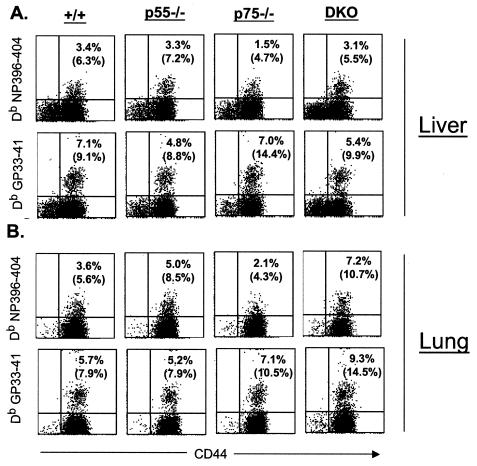
FIG. 5.
Virus-specific CD8 T cells in the nonlymphoid organs during a chronic LCMV infection. Eight days after infection with LCMV-clone 13, mononuclear cells were isolated from the liver and lung and stained with anti-CD8, anti-CD44, and MHC-I tetramers (Db) (loaded with CTL epitope peptides NP396 or GP33). The number of tetramer-binding CD8 T cells was quantitated by flow cytometry. The dot plots are gated on total CD8 T cells, and the numbers are the percentages of tetramer-binding CD8 T cells per total splenocytes. The numbers in parentheses are percentages of tetramer-binding CD8 T cells per total CD8 T cells.

