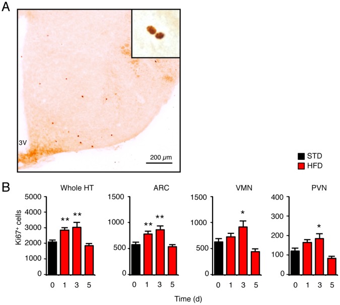
Figure 2. High-fat diet (HFD) transiently increased cell proliferation in the hypothalamus.
(A) Representative image of Ki67-positive proliferating cells immunodetected in the hypothalamus of control mice. (B) Quantification of Ki67-positive cells detected in the whole hypothalamus and in selected hypothalamic areas in mice fed either a standard or HFD for 1, 3 and 5 days. Basal amount of Ki67-expressing cells at day 0 corresponds to the value found in control mice fed a standard diet. Data are means ± SEM (n = 12 at day 0, and n = 5 at day 1, 3, and 5). Groups were compared using ANOVA followed if positive by Newman-Keuls post-hoc test. * and ** denotes p≤0.05 and p≤0.01, respectively. 3V: third ventricle; Arc: arcuate nucleus; HFD: high-fat diet; PVN: paraventricular nucleus; STD: standard diet; VMN: ventromedial nucleus.

