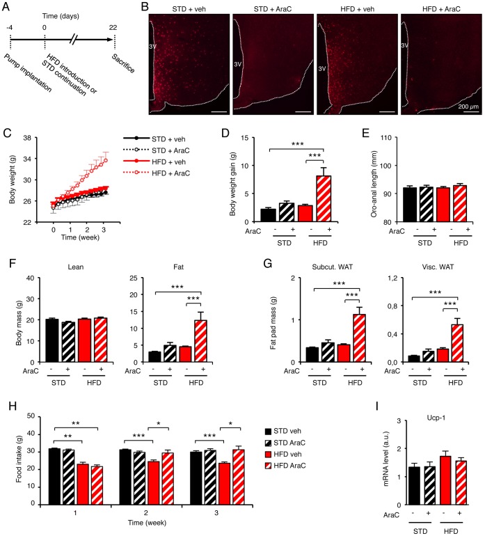
Figure 3. Blocking of the cell proliferation in the adult hypothalamus abolished the homeostatic feeding reponse to dietary fat and causes dramatic weight gain.
(A) Protocol used to inhibit cell proliferation in the brain. AraC-filled osmotic minipumps were implanted subcutaneously, and connected to the ventricular system to centrally deliver 40 µg/day AraC (at 0.25 µl/hr) for 3 weeks. Some mice also received BrdU 6 µg/day through the same route. Food intake and body weight were monitored during the time-course of the experiment, whereas body weigth gain, oro-anal length, adiposity, mass of fat depots and Ucp-1 expression were determined at the end of the treatment. (B) After 22 days, brains were analyzed by immunohistochemistry against BrdU to assess the efficiency of AraC treatment. (C) AraC increased body weight of HFD-fed mice from 8 days and after, in comparison to all other groups. (D–F) AraC dramatically increased body weight gain of mice fed with HFD for 3 weeks in comparison to all other groups. This effect did not affect the animal growth, but did increase their adiposity. (G) Masses of both subcutaneous (Subcut) and viscerous (Visc) fat pads were similarly increased by AraC. (H) From the second week after HFD introduction, AraC inhibited the continuation of the homeostatic reduction of food intake. (I) Levels of Ucp-1 mRNA in the brown adipose tissue assessed by RT-qPCR. No difference were found between groups, suggesting that the higher food intake in AraC-treated HFD-fed mice was not compensated by induced facultative thermogenesis. Data are means ± SEM (n = 12–15, except for fat pads weighing: n = 5 per group, and for Ucp-1 analysis: n = 8–9 per group). Groups were compared using ANOVA followed if positive by Newman-Keuls post-hoc test. *, **, *** denotes p≤0.05, p≤0.01, and p≤0.001 respectively. 3V: third ventricle. AraC: Arabinofuranosyl Cytosine; HFD: high-fat diet; STD: standard diet; veh: vehicle; WAT: white adipose tissue.

