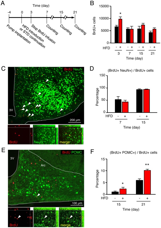
Figure 5. High-fat diet (HFD) did not deviate the neuronal fate rate, but incited new neurons to mature into anorectic POMC cells.
(A) Schematic presentation of the experimental design. Four days before HFD introduction, BrdU-filled osmotic minipumps were implanted subcutaneously and connected to ventricular system to centrally deliver 12 µg/day BrdU (at 0.5 µl/hr). Infusion was stopped at day 3 by cutting the cathether. Mice were kept for an additional 1 to 3-week period and then euthanized. Brain sections were examined after immunostaining of BrdU-positive neoformed cells. (B) HFD did not alter the survival rate at 3 weeks of BrdU-labeled neoformed cells in the hypothalamus in comparison to control mice. (C–D) HFD did not alter the differentiation rate into neurons in comparison to control mice, as assessed by double immunohistostaining against BrdU (in red) and NeuN, a neuronal marker (in green). (E–F) HFD increased the number of arcuate newborn POMC neurons, as assessed by double immunohistostaining against BrdU (in red) and POMC precursor (in green). Representative images were obtained from control mice at day 21. Data are means ± SEM (n = 5–6 per group). Groups were compared using unpaired t test. *, **, denotes p≤0.05 and p≤0.01, respectively. 3V: third ventricle; Arc: arcuate nucleus; HFD: high-fat diet.

