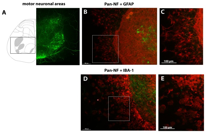
Figure 1. Motor neuron localization in organotypic spinal cord cultures.
(A) Displays the anatomy of spinal cord slices at lumbar level L5 modified from [46]. Areas containing motor neurons are highlighted in grey and with a box. On the left, the spinal cord is shown as illustration; on the right side a corresponding anti-pan-NF stained spinal cord slice after 7 days of cultivation is shown. (B and C) show anti-GFAP stained astroglia in organotypic spinal cord cultures after 7 DIVs (C) magnifies stained astroglia marked with a box in (B). (D and E) illustrate anti-IBA-1 stained microglia in these cultures. (E) magnifies stained microglia marked with a box in (D).

