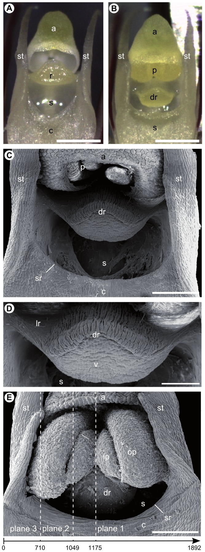
Figure 2. Apical gynostemium structures of Bulbophyllum bicoloratum viewed under the stereomicroscope (A, B) and scanning electron microscope (C–E).

(A) Isolated column of an outcrossing individual with erect rostellum at anthesis (without pollinia); (B) isolated column of a selfing individual with ‘displaced’ (suberect) rostellum at pre-anthesis (note, at anthesis, swollen pollina will obstruct the view on the rostellum; compare E); (C) column of a selfing individual with pollinia still in the anther at pre-anthesis; (D) close-up of the ‘displaced’ (suberect) rostellum (same as C) with the viscidium at its apex and cuticular folds on the upper (adaxial) side (except for the lateral sides); (E) column of a selfing individual after pollinia have been released from the anther onto the ‘displaced’ (suberect) rostellum. The swollen outer pollinia contact the (semi-) lateral rims of the stigmatic cavity (arrow). The dashed lines of planes 1–3 indicate approximate positions of longitudinal micro-CT sections shown in Figure 4A–C, respectively. The arrow below indicates the directionality of the complete scan (see Video S3), with numbers identifying scan-frames roughly corresponding to planes 1–3. Abbreviations: a, anther; c, column (gynostemium); dr, ‘displaced’ (suberect) rostellum (with cuticular folding); lr, lateral sides of ‘displaced’ (suberect) rostellum (devoid of cuticular folding); p, pollinia; ip, inner pollinium; op, outer pollinium; r, rostellum; s, stigmatic cavity; sr, rim of stigmatic cavity; st, stelidium; v, viscidium. Scale bars: (A, B) = 0.5 mm; (C, E) = 0.2 mm; (D) = 0.1 mm.
