
Figure 4. Effect of overexpression of FRMD7 on the actin cytoskeleton in NIH3T3 cells.
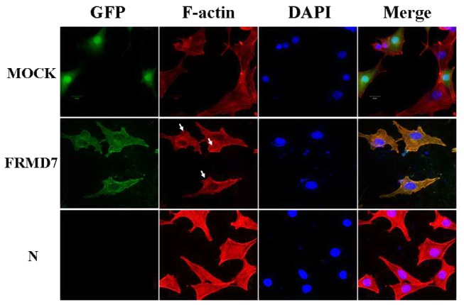
NIH3T3 cells transfected with mouse FRMD7-pEGFP-n1 (green) or the empty pEGFP-n1 vector (green) (Mock) were cultured for 24 hours and then cultured for 16 hours without FBS. F-actin was stained with TRITC-conjugated rhodamine-phalloidin (red). NIH3T3 cells transfected with the mouse full-length FRMD7 displayed some lamellipodia and ruffles, consistent with Rac1 activation. Normal NIH3T3 cells served as the control (N). Scale bars: 20 µm.