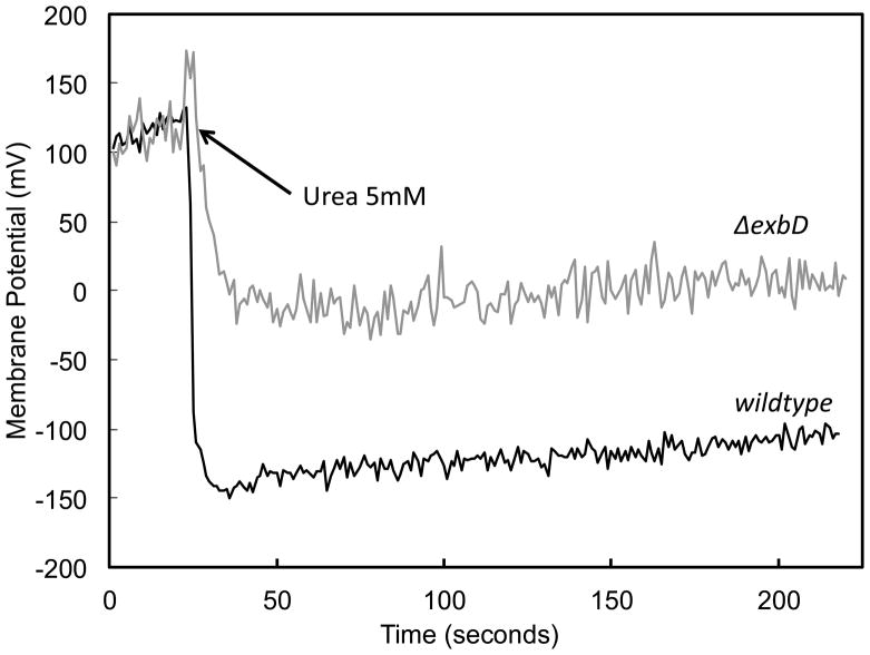
Figure 5.
Membrane potential measurement in the presence and absence of ExbD. DiSC3(5) was used to measure membrane potential at pH 3.0. In the wildtype (black line), with the addition of urea, there is a sustained increase in membrane potential, allowing maintenance of the proton motive force in the face of acidic medium pH. The ΔexbD (grey line) is unable to sustain an increase in membrane potential under the same conditions.

