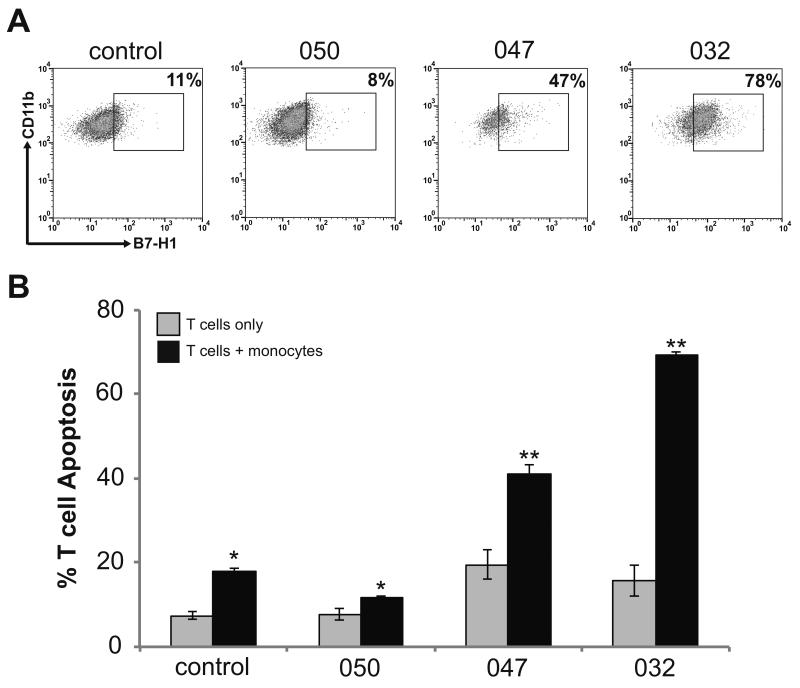
Figure 2. Peripheral blood monocytes from GBM patients induce autologous T cell apoptosis relative to B7-H1 expression.
(A) Gating demonstrating the percentage of B7-H1 positive monocytes isolated from peripheral blood of three GBM patients and a single non-glioma control patient. Monocytes were isolated from total peripheral blood leukocytes by CD14 positive selection. Cells were stained for flow cytometry and monocytes were identified by gating on CD45+/CD11b+ cells. This figure demonstrates the subgate of CD11b+/B7-H1+ cells. The cutoff for B7-H1 positivity was determined based on isotype control staining. (B) The percentage of T cells undergoing apoptosis when monocytes from patients in panel (A) were co-cultured with autologous activated T cells is shown. The percentage of T cell undergoing apoptosis was determined by co-staining cells for CD3 and Annexin V and analyzed by flow cytometry. Columns represent mean apoptotic T cell counts ± SEM from 3 independent trials. T cells co-cultured with monocytes demonstrated significantly increased apoptosis as compared to the same cells in culture alone (* p<0.05, ** p<0.01).

