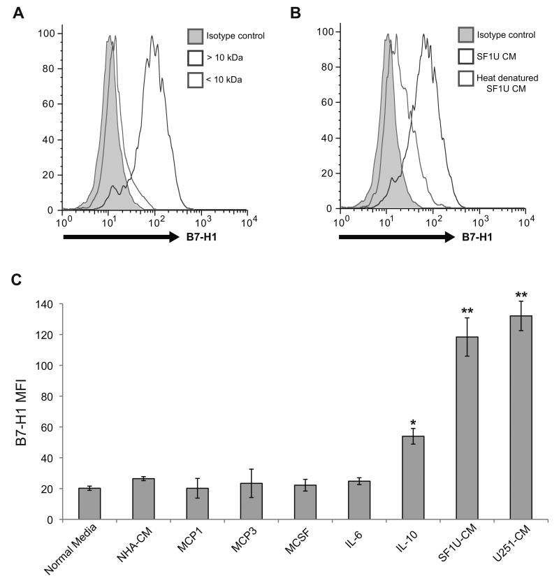
Figure 4. The glioma-derived B7-H1 inducing factor has properties consistent with a soluble protein.
(A) B7-H1 expression in normal monocytes treated for 24 hours with conditioned media from SF1U cells filtered to separate solutes with a molecular weight >10 kDa (dark gray) from solutes <10 kDa (light gray). The isotype control (gray shaded region) is also depicted. As shown, B7-H1 expression is significantly increased by a component in the > 10 kDa fraction. (B) B7-H1 expression in normal monocytes treated for 24 hours with unmodified conditioned media from SF1U cells (dark gray) or heat denatured media (light gray), demonstrating that heat denaturation eliminates the stimulatory effect of the conditioned media. (C) Mean fluorescence intensity of B7-H1 staining in normal monocytes treated with glioma-conditioned media or the following cytokines: MCP1 (200 ng/ml), MCP3 (200 ng/ml), M-CSF (50 ng/ml), IL-6 (10 ng/ml), IL-10 (10 ng/ml). Among the cytokines tested, only IL-10 stimulates a significant increase in B7-H1 expression (* p < 0.01). Compared to the response to IL-10, the increase seen with glioma-conditioned media stimulation was significantly greater (** p < 0.05). Columns represent mean fluorescence intensity ± SEM from 4 independent samples. Each sample was tested in triplicate and averaged as a single data point.

