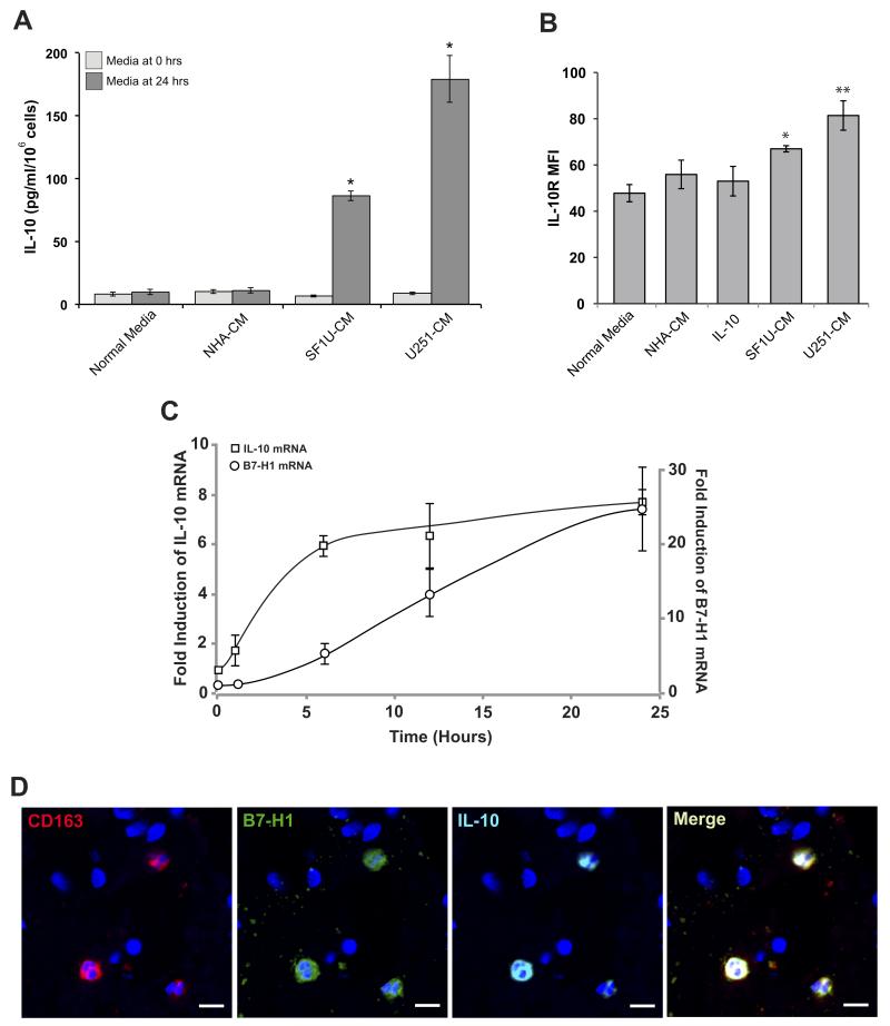
Figure 5. Glioma-derived soluble factor(s) enhance IL-10 signaling.
(A) Human IL-10 expression measured by ELISA from culture media of normal monocytes stimulated with glioma-conditioned media before and after 24 hours of culture. None of the conditioned media initially contained appreciable levels of IL-10. After 24 hours, IL-10 expression was significantly increased in cells stimulated with glioma-conditioned media compared to normal media (* p<0.01). Columns represent mean total IL-10 ± SEM from 3 independent samples. (B) IL-10 receptor expression was measured by flow cytometry in monocytes stimulated with glioma-conditioned media. Compared to unstimulated cells, treated monocytes had a modest but significant increase in IL-10R mean fluorescence intensity (* p < 0.05, ** p < 0.01). Columns represent mean fluorescence intensity ± SEM from 3 independent samples. Each sample was tested in triplicate and averaged as a single data point. (C) Time course of IL-10 and B7-H1 mRNA expression in normal monocytes stimulated with SF1U conditioned media. Transcript levels were measured at 0, 1, 6, 12, and 24 hours after stimulation using quantitative RT-PCR and normalized to 18S rRNA. Normalized expression is reported as a relative increase over initial expression. Each point represents mean relative expression ± SEM from 3 independent samples. (D) Representative section of GBM tissue with fluorescent immunestaining for CD163 (red), B7-H1 (green), IL-10 (cyan), and DAPI (blue) demonstrating co-localization of all three probes to the same subset of tumor-associated macrophages (scale bar = 10μm).

