
Figure 4.
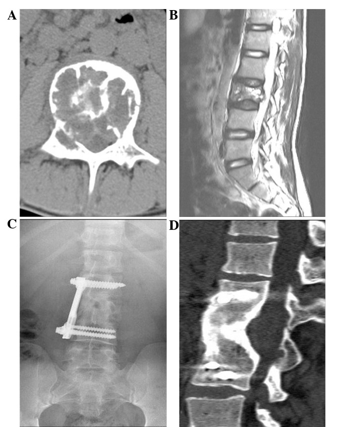
A 16-year-old male with a giant cell tumor affecting the L3 vertebra. (A) Axial CT scan of the L3 vertebra showing an osteolytic lesion in the vertebral body; (B) Sagittal T2-weighted MRI revealing invasion of the mass through the posterior wall of the vertebral body into the spinal canal; (C) Anteroposterior radiograph and (D) sagittal CT scan at 73 months follow-up, showing no recurrence. CT, computed tomography; MRI, magnetic resonance imaging.