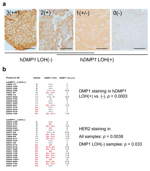
Figure 2. Histological grading of hDMP1 in human breast carcinoma.
a, human breast cancer tissues were stained with Dmp1-specifc RAX antibody (28) and the intensity of the nuclear staining was graded from 3(++), 2(+), 1(+/−), and 0 (negative). The scale bar is 100 μm.
b, correlation between LOH for hDMP1 and immunohistochemical grading of breast cancers. Breast cancer samples without LOH for hDMP1 showed significantly stronger nuclear signals for hDMP1. The hDMP1 signals were significantly higher in HER2 3+ or 2+ samples than in HER2 1+ or negative samples indicating the presence of the signaling pathway between HER2 and hDMP1 in breast cancers. Two different intensity values for hDMP1 indicate that the staining pattern for hDMP1 was mosaic; the average values (DMP1 scores) were used for statistical analyses.

