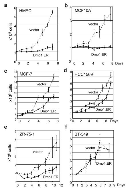
Figure 4. Proliferation assay of non-transformed human breast epithelial cells and breast carcinoma cell lines that overexpress Dmp1:ER.
(a) HMEC (human mammary epithelial cells); HER2low, ARF+, p53+
(b) MCF10A; HER2low, ARF−, p53+
(c) MCF7; HER2low, ARF−, p53+
(d) HCC1569; HER2++, ARF+, p53del
(e) ZR-75-1; HER2high, ARF+, p53+
(f) BT-549; HER2low, ARF+, p53mut
solid lines show the growth curves of Dmp1:ER virus-infected cells treated with 2 μM 4-HT, discontinuous lines show those of mock-infected cells with 4-HT.

