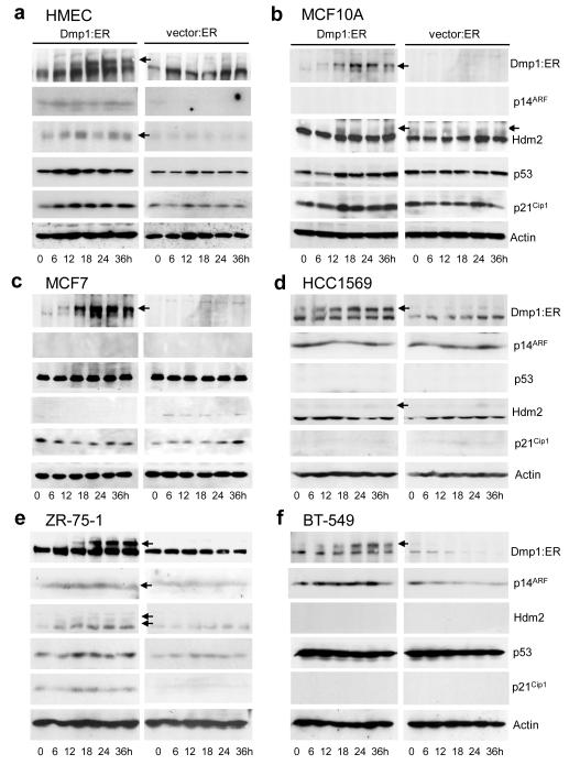
Figure 5. Western blotting analyses of (breast epithelial or cancer cells expressing activated Dmp1:ER or empty vector.
Lysate analyses were conducted by Western blotting with specific antibodies to Dmp1, p14ARF, p53, Hdm2, and p21CIP1. (a) HMEC, (b) MCF10A, (c) MCF7, (d) HCC1569, (e) ZR-75-1, and (f) BT-549 cells. Bottom axis shows hours after addition of 2 μM 4-HT.

