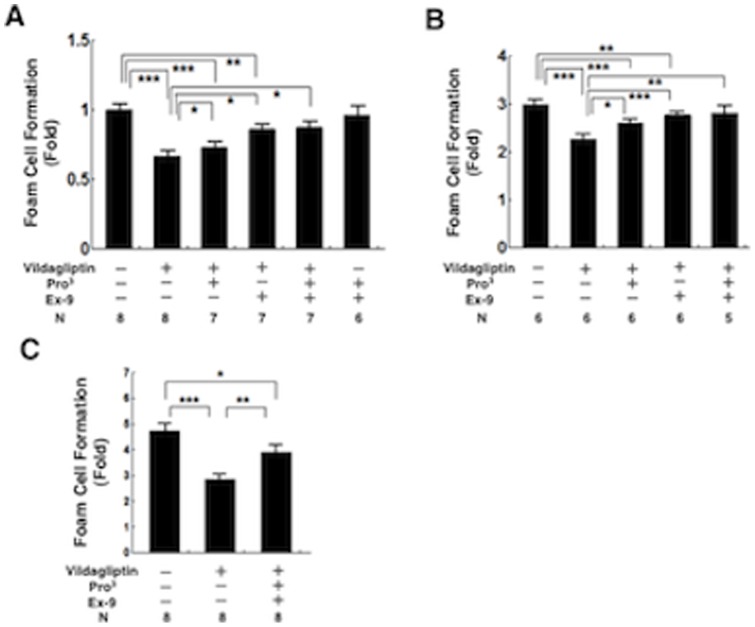
Figure 6. Foam cell formation in exudate peritoneal macrophages.
Exudate peritoneal cells were isolated from the treated nondiabetic Apoe −/− mice (a) or diabetic Apoe −/− mice (b) at 21 weeks of age, or db/db diabetic mice (c) at the age of 13 weeks, 4 days after an intraperitoneal injection of thioglycolate. Adherent macrophages were incubated for 18 hours with the RPMI-1640 medium containing 10 μg/ml oxLDL in the presence of 0.1 mmol/l [3H]oleate conjugated with bovine serum albumin. Cellular lipids were extracted and the radioactivity of the cholesterol [3H]oleate was determined by thin-layer chromatography. *P<0.05, **P<0.01, ***P<0.001.

