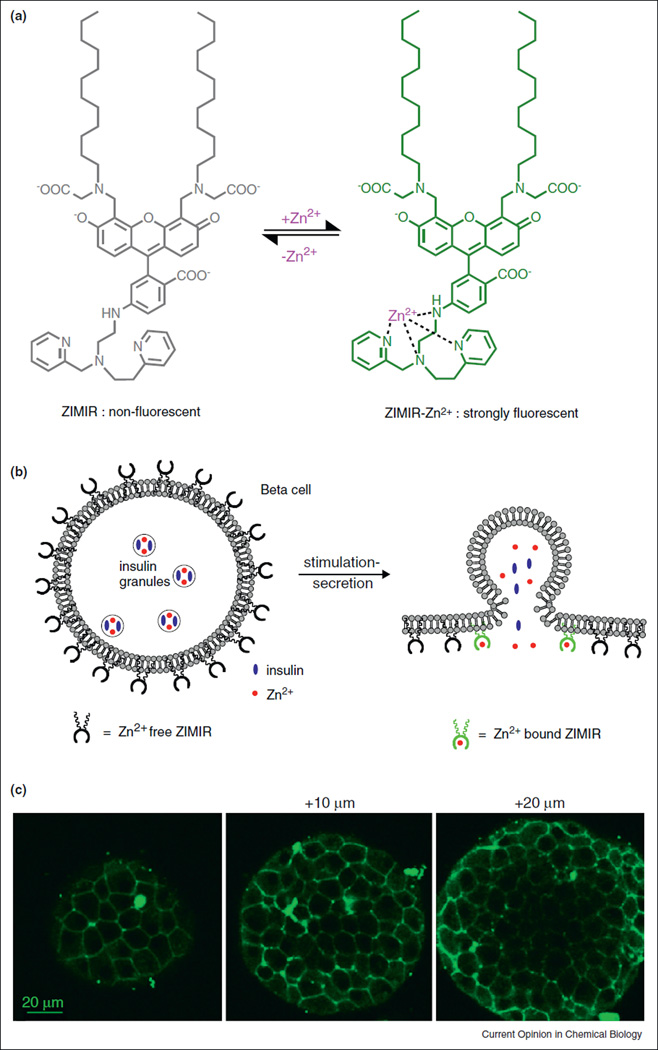
Figure 2.
A zinc indicator for monitoring induced exocytotic release (ZIMIR). (a) Chemical structure of ZIMIR in the Zn2+-free (nonfluorescent) and Zn2+-bound (strongly fluorescent) states. (b) Mode of action of ZIMIR for reporting local Zn2+ elevation at the membrane surface during exocytotic insulin granule fusion. The two lipophilic alkyl chains (wavy lines) anchor ZIMIR to the outer leaflet of the membrane lipid bilayer. (c) Confocal images of a ZIMIR-stained mouse pancreatic islet at three imaging depths. Adapted from [28].

