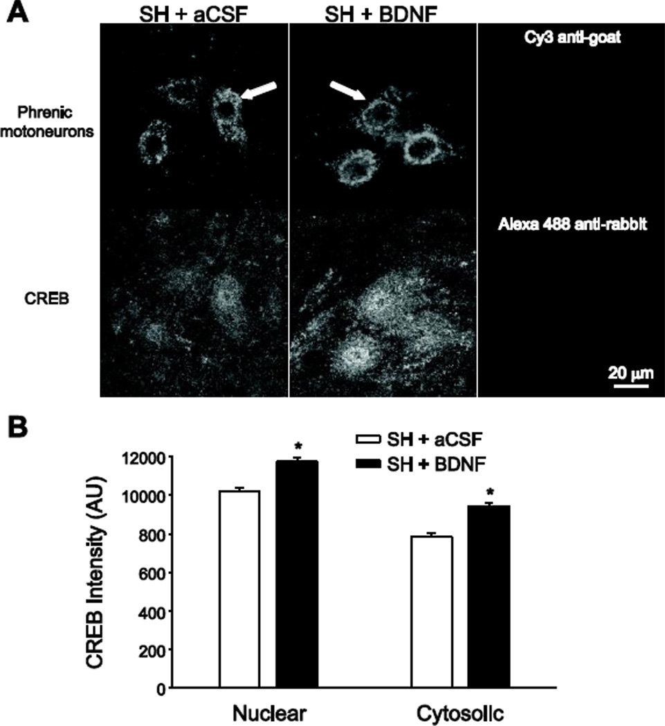
Figure 4.
Phrenic motoneuron immunoreactivity of the transcription factor cAMP response element-binding protein (CREB) following BDNF treatment. Simultaneous two-color imaging of phrenic motoneurons labeled retrogradely by intrapleural cholera toxin subunit B (CTB) injection (top panels in A) and CREB immunoreactivity (bottom panels in A) was used to quantify CREB immunofluorescence in the nucleus and cytosol of phrenic motoneurons (n=6/group). A) Representative images of CREB immunoreactivity in phrenic motoneurons at SH 7D in animals treated with intrathecal artificial cerebrospinal fluid (SH + aCSF; left panels) or intrathecal BDNF (SH + BDNF; middle panels). Images represent single confocal slices that were midnuclear for the phrenic motoneuron identified with an arrow. Right panels, representative images showing lack of immunofluorescence in tissues treated with secondary antibodies (but no primary antibody) and imaged at the selected acquisition parameters, which were constant throughout all samples. Bar, 20 µm for all images. B) The average fluorescence intensity (12-bit) was calculated in regions of interest in the cytoplasm and nucleus of CTBidentified phrenic motoneurons (n=421 and n=282 in the SH + aCSF and SH + BDNF groups, respectively), as well as in the surrounding background. Average values of CREB immunofluorescence for both nuclear and cytosolic regions (following background subtraction) are plotted. Both nuclear and cytosolic CREB immunoreactivity at SH 7D was greater following BDNF treatment compared to aCSF treatment (*, p < 0.001).

