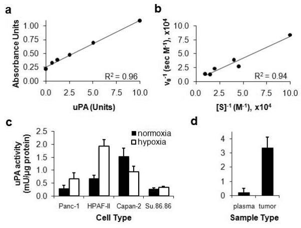
Figure 4.

Development of a pancreatic tumor model with high uPA activity. a) Total uPa activity was analyzed using the PAI Activity Assay Kit (ECM610 Kit, Millipore, Inc.) and a Synergy 2 microplate reader (Biotek Instruments Inc., Winooski, VA). Paired media samples without chromogenic substrate added were used for subtracting background absorbance. b) To ensure that enzyme concentration measured by this assay quantified enzyme activity, the initial rate of substrate cleavage was monitored as a function of substrate concentration and the results were analyzed using a Lineweaver-Burk plot. c) uPA activity was measured following two hours of incubation of the substrate with each cell type during in vitro studies conducted in normoxic and hypoxic conditions. The amount of uPa activity was normalized to total protein in each sample. Error bars represent the standard deviation of six trials for each cell type. d) The uPA activity was measured in blood plasma and homogenized tumor tissue. The amount of uPa activity in the samples was normalized to total protein in each sample. Error bars represent the standard deviation of results from four mice.
