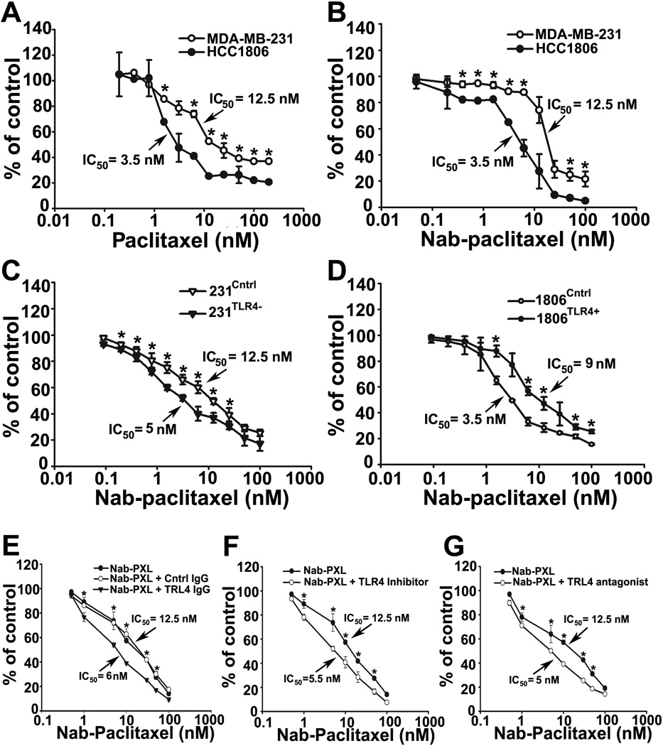
Figure 2. TLR4-positive and negative lines significantly differ in sensitivity to PXL.
MDA-MB-231 and HCC1806 cells were treated with PXL (A) or nab-PXL (B) at indicated concentrations (0–100nM) for 48hrs followed by calculating IC50. (C, D) 231Cntrl, 231TLR4− cells, 1806Cntrl and 1806TLR4+ cells were treated and analyzed as described under 2A. (E) MDA-MB-231 cells were pretreated with 4µg/ml of anti-TLR4 or control IgG for 2hrs followed by analysis described under 2A. (F, G) Cells were pretreated with a TLR4 inhibitor TAK-242 (10uM) or with an LPS antagonist LPS-EKUltra for 2hrs followed by analysis described under 2A. Data are presented as percent of viable cells ±S.D. from three experiments done in triplicate. The P-values represent * <0.05 vs. control as determined by Student’s paired t-test.

