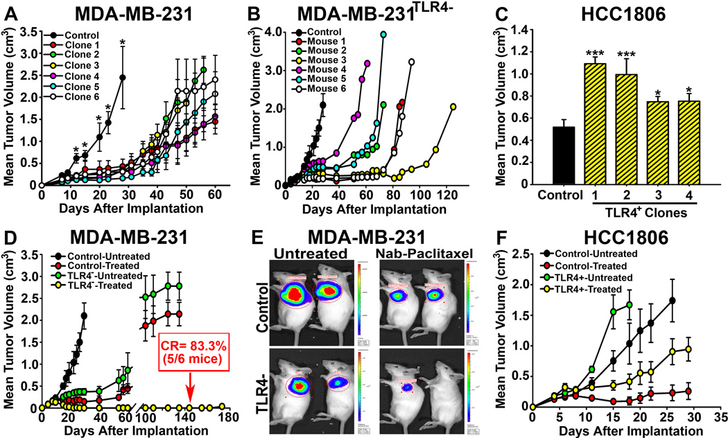
Figure 6. TLR4 determines sensitivity to nab-PXL in breast cancer modelsin vivo.
(A) The growth of 231TLR4− and control lines implanted in SCID mice was monitored twice weekly. Each point represents the mean tumor volume ±S.E and the asterisks indicate P-values >0.05. (B) Growth of a231TLR4− clone demonstrating significant delay in establishing tumor mass in all mice per group (n=6) compared with controls. Each line represents tumor growth in individual mouse. (C) Growth of HCC1806TLR4+ and HCC1806Cntrl tumorsin SCID mice. TLR4 significantly increased tumor growth in all clones with * and *** indicating P-values <0.05 and <0.001, respectively. (D) 231TLR4− and control tumors of 150 mm3 were treated with 10mg/kg of nab-PXL i.v. for 8 days. Five out 6 mice (83.3%) bearing 231TLR4− tumors (yellow circles) had complete response (CR) while 0% CRs was achieved in all other groups. (E) Bio-imaging of representative tumors from groups described in D.(F) Tumor growth of control and TLR4+overexpressing HCC1806 lines.

