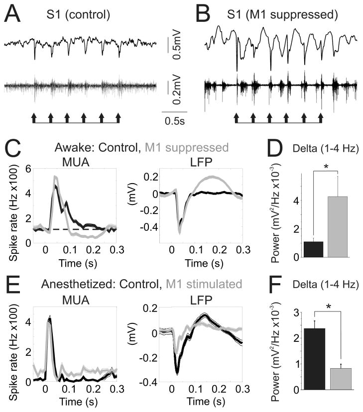
Figure 7. vM1 modulates S1 responses to simple sensory stimuli.
(A,B) Single trial S1 LFP (top) and MUA (middle) responses to brief (10 ms) whisker stimuli in waking mice, before (A) and during (B) focal vM1 suppression. Stimuli are indicated by the arrows below the traces. (C) Average MUA (left) and LFP (right) responses to whisker stimuli for control (black) and vM1 suppression (gray) conditions from one experiment. The dashed line (C, left) indicates baseline firing rates. (D) Population data, S1 LFP delta power during sensory responses in control (black) and vM1 suppression (gray) conditions. (E–F) Experiments in anesthetized mice, pairing brief deflections of the principal whisker with vM1 stimulation. (E) Average MUA (left) and LFP (right) responses to whisker stimuli for control (black) and vM1 stimulation (gray) trials from one experiment. (F) Population data, S1 LFP delta power during sensory responses in control (black) and vM1 stimulation (gray) conditions. *, p<0.05.

