Fig. 2.
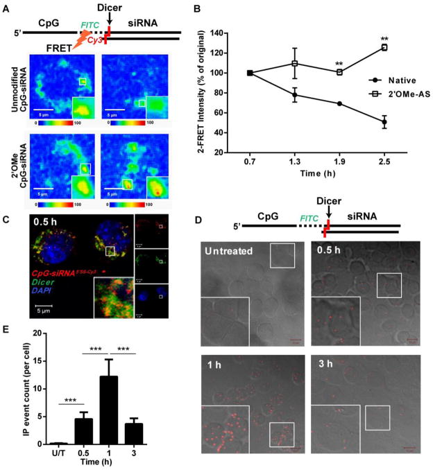
CpG-siRNA conjugates are uncoupled in the presence of Dicer endonuclease shortly after internalization into RAW 264.7 cells. (A) Separation of the CpG and siRNA parts the CpGFITC-Stat3 siRNA5′AS-Cy3 conjugate was measured by decreasing FRET effect using time-lapse confocal microscopy. The nuclease-resistant conjugate with completely 2′OMe-modified both strands of the siRNA was used as a negative control. Shown are representative results from one of three independent experiments. Scale bars: 5 μm. (B) Quantification of average FRET intensities from three independent experiments using unmodified and 2′OMe-modified conjugates. Shown are means ± SEM. (C) CpG-siRNA5′SS-Cy3 colocalizes with Dicer within 0.5 h after uptake as assessed by confocal microscopy on fixed RAW 264.7 macrophages. (D, E) Dicer transiently interacts with CpG-siRNA shortly after endocytosis of the conjugate. Cells were incubated with CpGFITC-siRNA for indicated times, fixed, permeabilized and the interaction between CpGFITC-siRNA and Dicer was detected by in situ PLA using FITC- and Dicer-specific antibodies. (D) Shown are confocal microscopy images (Z stack projections; Scale bars: 10 μm) and (E) the average counts of PLA events per cell for indicated times, means ± SEM.

