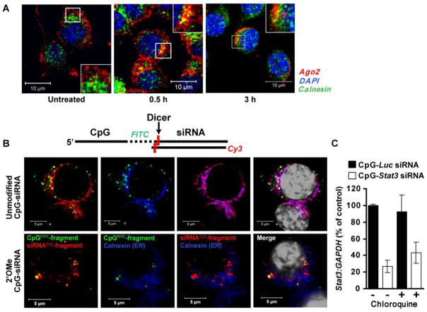
Fig. 3.
Diced siRNA escapes from EE to endoplasmic reticulum. (A) Treatment with CpG-siRNA induces partial translocation of Ago2 to the ER. Shown are confocal microscopy images acquired from primary mouse bone-marrow derived macrophages. Scale bars: 10 μm. (B) The intracellular localization of both parts of CpG-siRNA after endosomal processing. Unmodified (top row) and 2′OMe-modified (bottom row) CpGFITC–Stat3 siRNA5′SS-Cy3 were added to cultured RAW 264.7 cells. After 3 h, cells were fixed, stained with anti-calnexin antibody (ER marker) plus DAPI (nuclear staining; shown in white) and analyzed by confocal microscopy. Scale bars: 5 μm. (C) Disruption of late endosomes by chloroquine does not affect target gene silencing by CpG-Stat3 siRNA. Stat3 gene silencing was measured using qPCR in 264.7 cells treated with CpG-Stat3 siRNA and CpG-Luc-siRNA (negative control) with or without chloroquine (100 μM), inhibitor of endosomal maturation, as indicated. Shown are representative results from one of two independent experiments; mean ± SEM.

