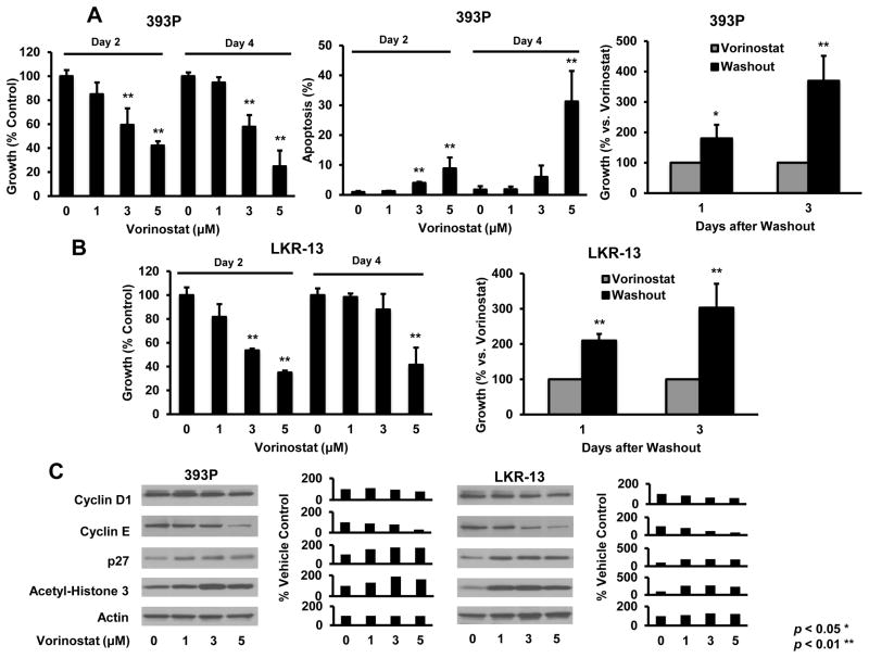
Fig. 2.
Vorinostat effects in murine 393P (KrasLA1/+; p53R172HΔG/+) and LKR-13 (KrasLA2/WT) transgenic lung cancer cell lines. (A) Vorinostat inhibited growth in a dose-and time-dependent manner and increased apoptosis in 393P cells. Effects were antagonized by vorinostat washout. (B) Similar proliferation, apoptosis (data not shown) and washout effects occurred in LKR-13 cells. Vorinostat treatments decreased cyclin D1 and cyclin E proteins, but increased p27 and acetyl-histone H3 protein levels in 393P (left panel C) and LKR-13 (right panel C) cells. Respective signal intensities are quantified in the right panels). Symbols * and ** revealed significant changes P < 0.05 and P < 0.01, respectively.

