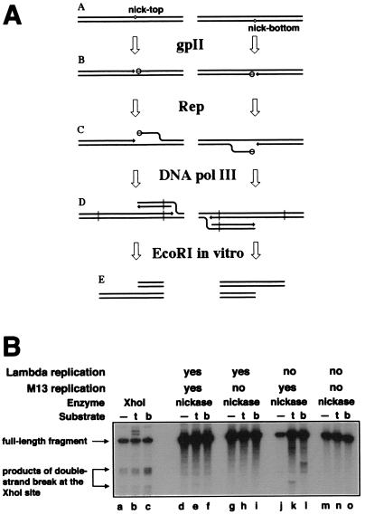
Figure 3.
The interplay between λ and M13 DNA replication. (A) Sigma replication in Rep+ cells is predicted to generate the “half-break” pattern at the nicking sites. 3′-ends are indicated by arrows. (A) The substrate chromosomes carrying nicking sites at the same location, either in the top or in the bottom strand. (B) The nicking sites are nicked by gpII (open circles attached to the 5′-sides of the nicks). (C) The Rep helicase unwinds the 5′-ends of the nicks, attracting replisomes to the replication fork structures. (D) Sigma replication from the nicks. (E) Restriction cutting in vitro (at sites indicated by vertical lines in D) reveals the “half-break” pattern. (B) Interference between λ DNA replication and nicking by the wild-type nicking enzyme. Phages MMS2660, λAK2, or λAK3, indicated in the entry “substrate” as “—”, “t,” or “b,” respectively, were infected at moi = 6 into the strains described below; the cells were incubated as indicated, and the phage DNA was extracted and analyzed by restriction digestion with EcoRI and blot hybridization with probe 1. Molecular weight markers for double-strand break at the natural XhoI site were generated in vivo (lanes a–c). The strains and conditions are as follows: lanes a–c, recB268 pK107, 10 min at 28°C; lanes d–f, recB270 recC271 pCL475, 30 min at 42°C; lanes g–i, Δrep∷cam recB270 recC271 pCL475, 30 min at 42°C; lanes j–l, recB270 recC271 pCL475, incubated for 30 min at 42°C before phage injection and 60 min at 28°C after phage injection; lanes m–o, Δrep∷cam recB270 recC271 pCL475, incubated for 30 min at 42°C before phage injection and 60 min at 28°C after phage injection.

