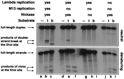
Figure 5.
Replication-induced double-strand breaks at persistent nicks at the XhoI-site on the other side of the λ replication origin. Phages MMS2663, λAK4, or λAK5, indicated in the entry “substrate” as “—”, “t,” or “b,” respectively, were infected at moi = 6 into the strains described below; the cells were incubated at 28°C for 80 min, and the phage DNA was extracted and analyzed by restriction digestion with PstI + NcoI and blot hybridization with probe 2 under both neutral (Upper gel) and denaturing (Bottom gel) conditions. Molecular weight markers for “half-breaks” at the XhoI site were generated in vivo (lanes a–c). The strains and conditions are as follows: lanes a–c, recB268 pK133 + IPTG; lanes d–f, Δrep∷cam recD1011 pK125 + IPTG; lanes g–i, Δrep∷cam recD1011 pK125, no induction; lanes j–l, Su− Δrep∷cam recC1010 pK125 + IPTG. To compensate for the smaller amount of λ DNA because of the inhibition of λ replication, 25 times more total DNA has been loaded in the case of Su− samples (lanes j–l).

