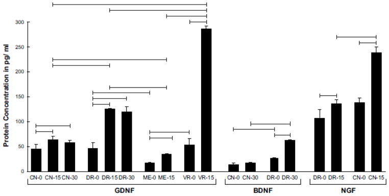
Figure 6.

Concentrations of GDNF, BDNF, and NGF protein in normal cutaneous nerve, dorsal root, ventral root, and muscle nerve, and in selected populations of denervated Schwann cells. Horizontal bars linking different groups indicate a significant difference in concentration (p< 0.05). GDNF was upregulated prominently by DR and VR, and the resulting protein concentrations were significantly higher in these structures than in their peripheral extensions, CN and ME. The BDNF profile is consistent with the PCR findings after 30 days of denervation, with expression in CN returning to baseline and that in DR peaking. The increased pace of NGF upregulation in CN when compared with DR was also reflected in protein concentrations.
