
Fig. 4.
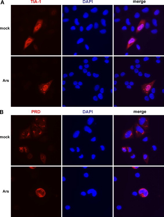
Stress granule formation following expression of a dominant negative version of TIA-1. SK-N-SH cells were transfected with HA-tagged TIA-1 or TIA-1-PRD (or empty vector, not shown) and either mock-stressed (top panels in A and B) or stressed with sodium arsenite (final concentration 0.5 mM; bottom panels in A and B) for 30 min at 37 °C. Cells were washed and fixed, incubated with an anti-HA monoclonal antibody, and subsequently AlexaFluor594 goat anti-mouse secondary antibody. Cells were examined for the formation of stress granules via fluorescence microscopy; nuclei were identified by DAPI staining.