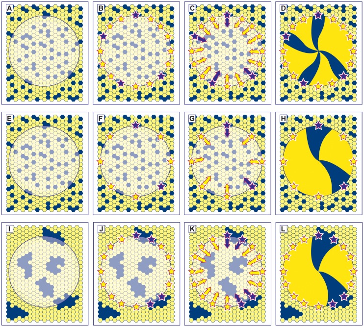
Figure 1. Alternative hypotheses to explain the reduction in corrected stripe numbers in Pax6+/− mosaic corneas.
(A–D) Normal development: The developing mosaic surface ectoderm (A) comprises two genetically marked cell populations (shown as blue and yellow hexagons) and the future corneal epithelium is shown as a disk with the limbus at its periphery. LESCs are specified (shown as stars in B) from a pool of cells in the surface ectoderm and become active postnatally (C). Centripetal movement of daughter TACs forms radial stripe patterns in the adult corneal epithelium (D). (E–H) Hypothesis 1– reduced Pax6+/− LESC numbers: If fewer LESCs are specified in the Pax6+/− ocular surface (F) then individual clones of corneal epithelial cells produced by each LESC will colonise a larger sector of the cornea, forming fewer, wider stripes (H). A similar result is expected if normal numbers of LESCs are specified but some fail to survive. (I–L) Hypothesis 2– reduced cell mixing during Pax6+/− development: If there is less mixing of the two genetically marked cell populations in the Pax6+/− mosaic surface ectoderm during development, cells will be grouped into larger coherent clones to form a coarse-grained mosaic pattern (I). There is a higher probability that two adjacent stem cells belong to the same population (e.g. adjacent blue stem cells in J) so wider stripes are produced. Although, in this case, the distribution of LESCs around the circumference will be non-random, the distribution of LESC clones should still be random. The corrected stripe number described in the text is expected to be proportional to the number of LESC clones but the number of LESCs per clone may vary as shown (e.g. compare H and L).

