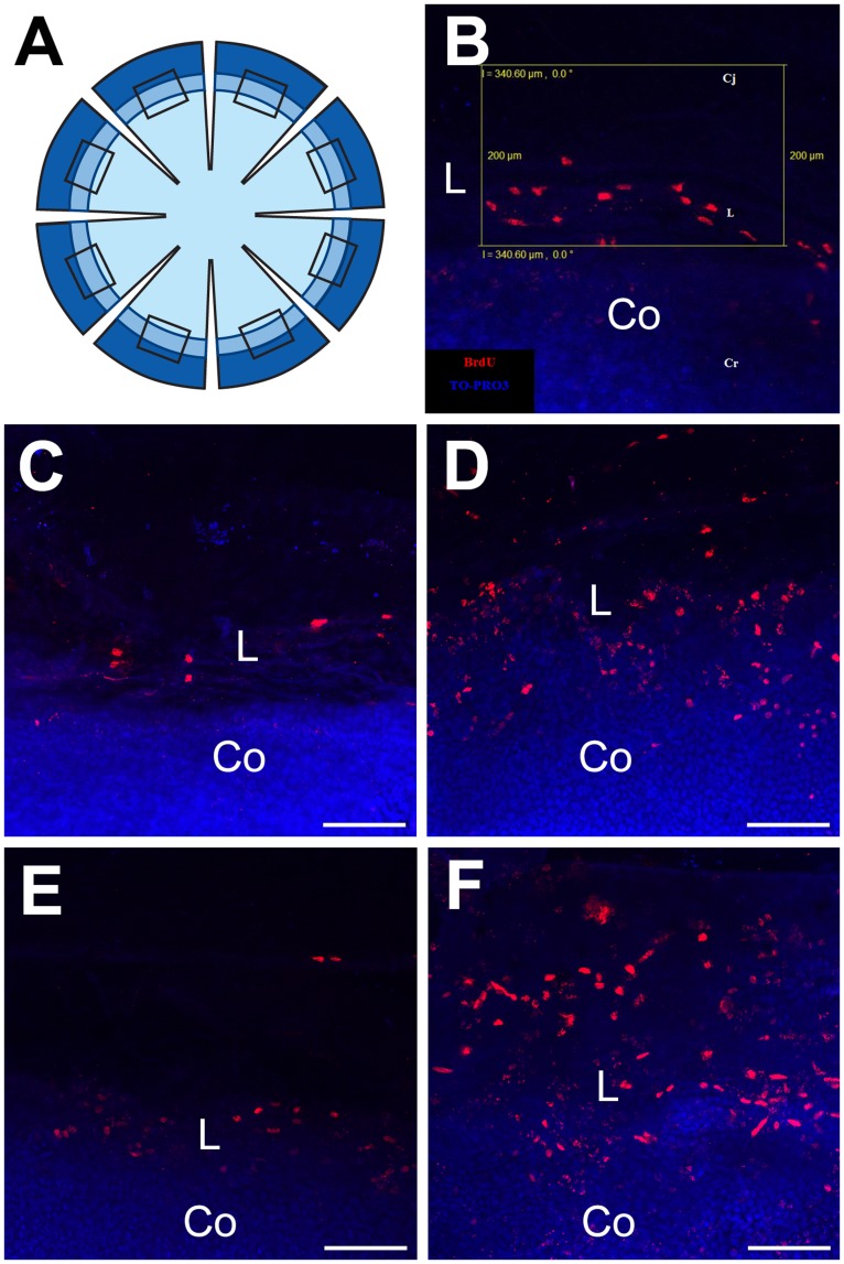
Figure 6. Identification of label-retaining cells in the limbal region of the WT and Pax6+/− ocular surface epithelium.
(A) Diagram showing the 8 radial cuts in a corneal button, shaded to represent the cornea (lightest), limbus (intermediate) and conjunctiva (darkest). The rectangles show the location of the sampling boxes (one per sector but not to scale). (B) Rectangular 340×200 µm sampling box (yellow outline), used to count LRCs, superimposed on the limbal region of an image of BrdU-labelled nuclei (red) counterstained with TO-PRO3 iodide (blue) in a whole mount flattened corneal button with associated conjunctival tissue. (C–F) Examples of BrdU label-retaining cells in the limbal region after 1-week BrdU exposure and 10 week chase period in (C) 15-week old WT, (D) 15-week old Pax6+/−, (E) 30-week old WT and (F) 30-week old Pax6+/−. Pixel resolution: 1024×1024. Abbreviations: Cj: Conjunctiva; L: Limbus; Co: Cornea. Red immunofluorescence: BrdU; Blue: TO-PRO3 iodide counterstain. Scale bars are 100 µm.

