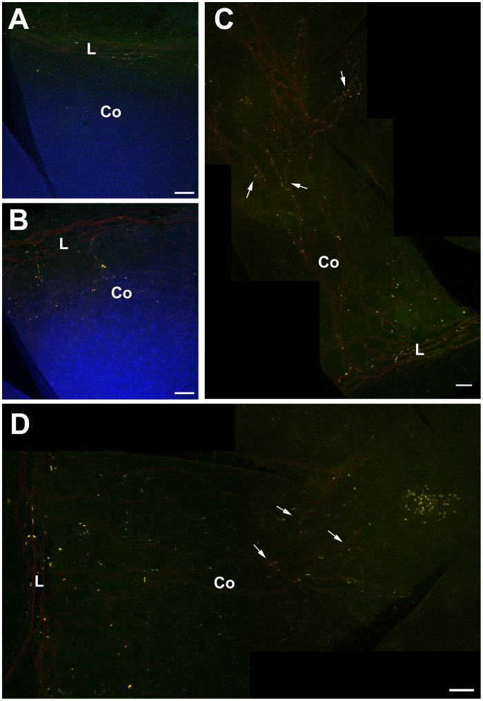
Figure 8. Association of BrdU label-retaining cells with blood vessels in Pax6+/− corneas.
Double immunofluorescent detection of BrdU (yellow) and CD31-positive blood vessels (red) in WT and Pax6+/− mice. (A) Flattened WT cornea showing BrdU-positive, label-retaining cells (LRCs) and CD31-positive blood vessels in the limbus. (B) Flattened Pax6+/− cornea demonstrating CD31-positive blood vessels extending from the limbus into the cornea and BrdU LRCs in the cornea. (C, D) Montages of flattened Pax6+/− corneas showing CD31-positive blood vessels and BrdU LRCs even in the central cornea. Arrows show blood vessels with adjacent BrdU-positive cells (LRCs). For demonstration purposes the counterstain channel was deactivated. Abbreviations: L: Limbus; Co: Cornea; Yellow immunofluorescence: BrdU. Red immunofluorescence: CD31; Blue: TO-PRO3 iodide counterstain. Scale bars are 100 µm.

