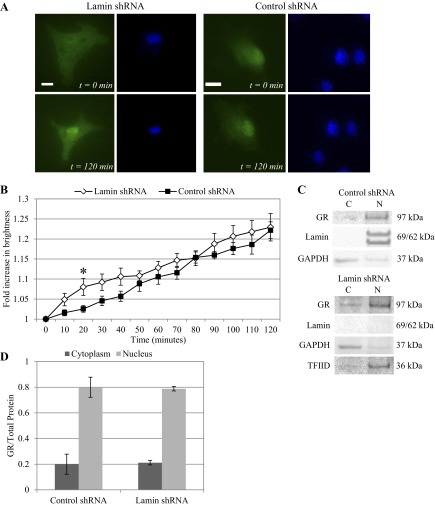
Fig. 3.
Lamin deficiency did not prevent GFP-GR nuclear movement under shear stress at 10 dyn/cm2. A: live cell imaging of GFP-GR in BAECs transfected with lamin shRNA showed increasingly nuclear localized GR after 120 min of shear stress at 10 dyn/cm2 (left). Similar GFP-GR movement was also observed in cells with control shRNA (right). Nuclei were labeled with Hoechst stain. Images were taken at ×40 magnification. Scale bar = 15 μm. B: quantitative image analysis of GFP-GR subcellular movement shows a 22.9 ± 3.4% and 22.2 ± 2.1% increase in nuclear brightness after 2 h of shearing in BAECs with lamin (n = 7) and control (n = 14) shRNA respectively. *P < 0.05 at t = 20 min for control vs. lamin shRNA-treated BAEC. C: Western blot of endogenous GR shows similar increase of GR in the nuclear (N) protein fractions compared with cytoplasmic (C) in both cell types after 120 min of shear. Lamin A/C and TFIID served as the nuclear protein controls, and GAPDH was used as control for cytoplasmic protein. D: quantitative analysis of cytoplasmic and nuclear GR fractions based on repeated Western blots shows a significant increase of GR in the nuclear fraction compared with cytoplasmic proteins (n = 3).

