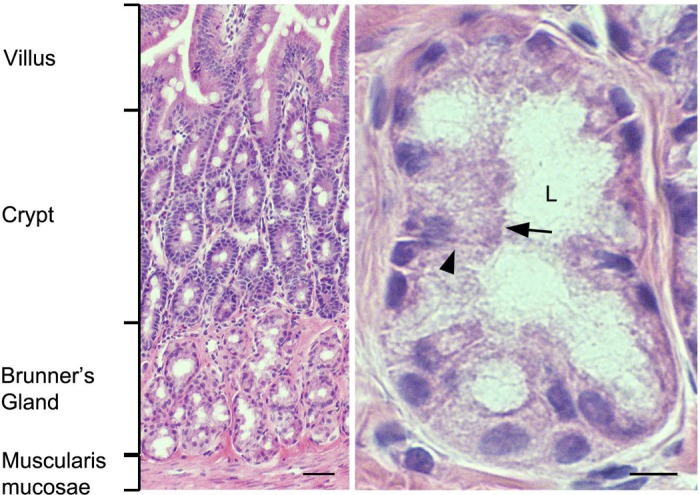
Fig. 1.

Histological appearance of rat duodenum Brunner's glands. Hematoxylin and eosin-stained sections from rat proximal duodenum. Low-magnification image (left) shows location of Brunner's glands relative to the crypt-villus axis. High-magnification image (right) of representative Brunner's gland. Black arrow indicates apical membrane; black arrowhead indicates basolateral membrane. L, lumen. Scale bar = 10 or 100 μm.
