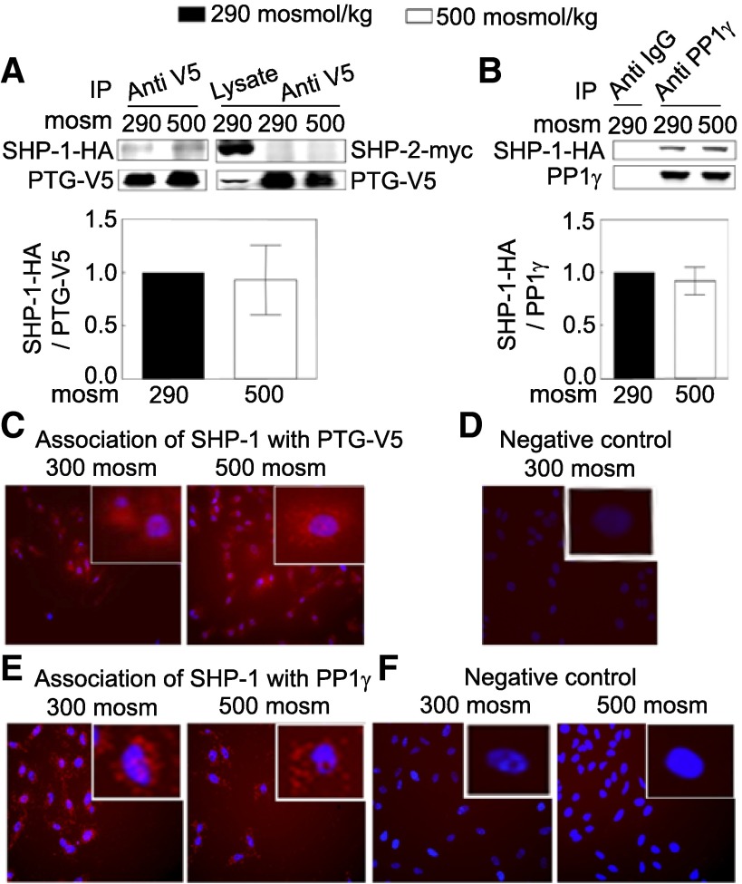
Fig. 5.
A: PTG-V5 is physically associated with SHP-1 but not SHP-2, and the association is not affected by high NaCl. HEK293 cells were co-transfected at 290 mosM with plasmids coding for PTG-V5 and with SHP-1-HA or SHP-2-myc for 48 h. Then, osmolality was increased to 500 mosM (NaCl added) or left at 290 mosM for 30 min before cell lysates were prepared. The lysates were immunoprecipitated with mouse anti-V5 antibody, and Western blots were probed with rabbit anti SHP-1 or SHP-2 antibody (n = 3). B: PP1γ is physically associated with SHP-1, and the association is not affected by high NaCl as in A, except that cells were transfected with the plasmid coding for SHP-1-HA but not for SHP-2-myc. The lysates were immunoprecipitated with the goat anti-PP1γ antibody, and Western blots were probed with rabbit anti-SHP-1 antibody (n = 3). C–F: Duolink in situ assays. Red stain indicates protein-protein association. Blue stain indicates nuclei, detected by DAPI. C: SHP-1 associates with PTG-V5 at 300 and 500 mosM. HeLa cells were transfected with the plasmid coding for PTG-V5 for 24 h. Cells were plated on a chamber slide (NUNC) for an additional 24 h, and then osmolality was increased to 500 mosM (NaCl added) or left at 300 mosM for 30 min before Duolink in situ assay. Mouse anti-V5 antibody (1:250) and rabbit anti-SHP-1 (1:50) were used. D: negative control. Same as in C, except the HeLa cells were not transfected with the PTG-V5 plasmid. E: SHP-1 associates with native PP1γ at 300 and 500 mosM. Nontransfected HeLa cells were incubated with the goat anti-PP1γ (1:50) and rabbit anti-SHP-1 (1:50) for the Duolink in situ assay. F: negative control. Same as in E, except no anti-SHP-1 antibody was included in the Duolink in situ assay. (All Duolink assay results are representative of two independent experiments).

