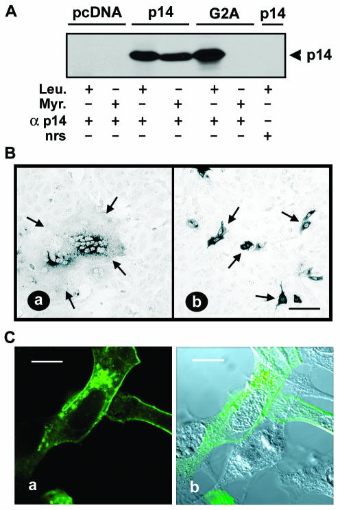
FIG. 7.
N-terminal myristylation of p14 is essential for cell fusion. (A) 2HAC-tagged p14-, p14-G2A-, and pcDNA3-transfected Vero cells were labeled with either [3H]myristic acid (Myr.) or [3H]leucine (Leu.) and immunoprecipitated with anti-p14 polyclonal antiserum (α p14) or normal rabbit serum (nrs). Precipitates were fractionated by SDS-PAGE and radiolabeled p14 was detected by fluorography. (B) Vero cells transfected with either 2HAC-tagged p14 (a) or p14-G2A (b) were fixed with methanol at 18 h posttransfection and immunostained with anti-p14 polyclonal antiserum and an alkaline phosphatase-conjugated secondary antibody. Arrows indicate the limits of an antigen-positive syncytial focus (a) or individual antigen-positive cells expressing the fusion-negative p14-G2A construct (b). Scale bar = 100 μm. (C) QM5 cells transfected with p14-G2A were permeabilized with methanol and immunostained at 9 h posttransfection with anti-p14 polyclonal antiserum and FITC-conjugated secondary antibody. Fluorescence microscopy (a) revealed punctate, perinuclear intracellular staining of p14-G2A and a ring surface fluorescence characteristic of authentic p14. The corresponding DIC image overlaid with the fluorescent image is also shown (b). Scale bar = 10 μm.

