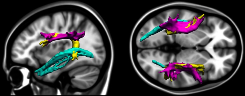
Figure 1.
Illustration of the tracts of interest. Tracts of interest were estimated in each individual's native diffusion space, extracted from an example participant, and registered to MNI template space for visualization here (sagittal view on the left, axial on the right). The ILF (cyan) spans the occipital and temporal cortices. The SLFp (magenta) connects frontal and parietal regions. The other component of the SLF is the arcuate fasciculus (yellow), which departs from the main SLF tract to branch into temporal cortices and is classically posited to facilitate communication between Broca's and Wernicke's areas.

