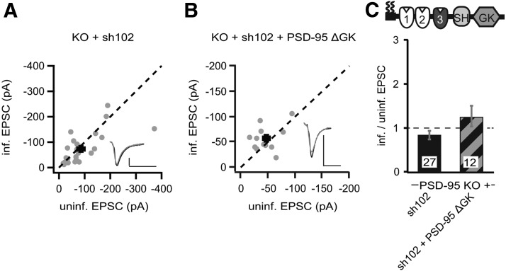
Figure 8.
p95ΔGK-mediated enhancement of AMPAR EPSCs in PSD-95 KO mice was dependent on increased SAP102 levels. A, B, Amplitude peaks of AMPAR EPSCs of neurons expressing sh102 (A) or sh102 + p95ΔGK (B) are plotted against those of simultaneously recorded uninfected neighboring neurons in PSD-95 KO hippocampal slice cultures. C, Summary of AMPAR EPSC ratio of infected and uninfected pairs. Schematic representation of PSD-95 with color-coded domains, matching the bar colors. Number of pairs (n) indicated in the foot of the graph. Calibration: 50 pA, 25 ms.

