Figure 6.
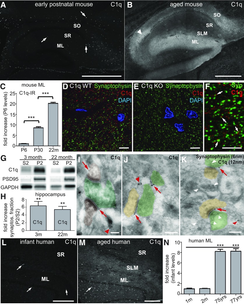
C1q immunoreactivity dramatically increases with age in hippocampal synaptic layers and at synapses. A, B, Immunohistochemistry analysis confirmed comparably low levels of C1q immunoreactivity (arrows) in the hippocampus of P6 mice (A) but dramatically increased levels in all synaptic layers of the 24-month-old murine hippocampus (B): stratum oriens (SO), stratum radiatum (SR), stratum lacunosum moleculare (SLM), dentate gyrus ML. Very strong C1q immunoreactivity was also detected in the neuropil surrounding the CA2 cell bodies (arrowhead). C, Quantification of C1q immunoreactivity in the ML from early postnatal to old aged mice confirmed that C1q immunoreactivity dramatically increases with aging in the murine ML. C1q signals, normalized to P6 levels (n = 3 brains per age; statistical significance: P30 vs P6: p < 0.001; 22 months vs P30: p < 0.0001). D, E, Anti-C1q immunohistochemistry signals (rabbit anti-mouse C1q monoclonal antibody) visualized by confocal microscopy were specific. C1q immunoreactivity was only detected in the adult wild-type dentate gyrus molecular layer (D) but not in the corresponding tissue from C1q-deficient littermate mice (E). F, High resolution confocal microscopy revealed that C1q immunoreactivity in the ML was detected in close proximity to synaptic terminals (synaptophysin immunoreactivity, arrows). G, Subcellular fractionation of murine hippocampus homogenate revealed that C1q (detected with a polyclonal anti-C1q antibody) was enriched in the crude synaptosomal fractions (P2) from both adult and aged mice, similar to the synaptic protein PSD95, compared with the supernatant fraction (S2; representative images; GAPDH loading control). H, Quantification of the hippocampal subcellular fractionation analysis confirmed that C1q was enriched in the crude synaptosomal fraction (P2) from both 3-month-old (p = 0.0091) and 22-month-old (p = 0.0055) mice (n = 4 animals/age), compared with the counter-fraction S2. I–K, Single-section cryo-immuno electron microscopy confirmed that C1q immunoreactivity (12 nm gold particles) was detected in close proximity to the extracellular side of presynaptic- (pseudo-colored in green) or postsynaptic- (pseudo-colored in brown) elements (red arrows) and associated with unidentifiable structures (red arrowheads). This single-section image analysis did not allow clear identification of all elements visible in each section. It was therefore impossible to classify C1q immunoreactivity associated with unidentified elements as synaptic or nonsynaptic. K, Dual-immunogold analysis in the dentate gyrus molecular layer of aged mice. C1q immunoreactivity (12 nm gold particles, arrows) was detected in close proximity to both presynaptic (green) and postsynaptic (brown) elements. In contrast, synaptophysin immunoreactivity (6 nm gold particles, white arrowheads) was detected inside clearly identifiable presynaptic terminals as well as associated with unidentifiable structures, likely reflecting presynaptic terminals. L, M, C1q immunoreactivity dramatically increased with age in synaptic layers of the human hippocampus. L, C1q immunoreactivity in the human infant (2-month-old) was restricted to blood vessels (arrows), indicating serum-derived C1q in this nonperfused tissue, whereas it was particularly enriched in the SLM and ML of aged human donors (M; 75 years old). N, Quantification of C1q immunoreactivity in the ML from available infant and old aged human donor tissue confirmed that C1q immunoreactivity dramatically increased with aging in the human ML. C1q signals, normalized to 1 month levels (n = 4 slices per donor; statistical significance: 1 vs 2 months: p < 0.79; 1 month vs 75 years: p < 0.0001; 1 month vs 77 years: p < 0.0001). **p < 0.01, ***p < 0.001. Values and error bars represent mean ± SEM. Scale bars: A, B, L, M, 500 μm; D–F, 5 μm; I–K, 200 nm.

