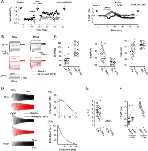
Figure 2.
Group 1 mGluR activation produces long-lasting changes in the subthreshold membrane properties of CPn but not COM neurons. A, RMP or RN at −65 mV was monitored over time in CPn and COM neurons. The application of DHPG produced a long-term hyperpolarization of RMP and an increase in RN in CPn neurons. In contrast, in COM neurons, there was only a transient depolarization of RMP. Arrows indicate the relevant times at which measurements were made for subsequent panels. The sADP was measured during the 10 min post-DHPG time point. B, Sample voltage responses to a family of current injections before and 30 min after a 10 min application of DHPG. RN, sag, and rebound were calculated from these voltage responses. C, DHPG produced an increase in RN and a decrease in sag and rebound in CPn neurons. There were no changes in these measurements in COM neurons. *p < 0.001 (paired t test). D, Sample voltage responses to a chirp stimulus current injection. These responses were used to create the impedance amplitude profiles (ZAPs) shown at the right. The peak of the ZAP is the resonant frequency. E, DHPG produced a decrease in resonance in CPn neurons, whereas resonance in COM neurons remained 1 Hz. All Ih-related measurements were made at −65 mV. *p < 0.001 (paired t test). F, DHPG produced a long-lasting hyperpolarization of the RMP in CPn neurons and a transient depolarization of the RMP in COM neurons. The peak change refers to the peak change in RMP at any time during the DHPG application and washout. *p < 0.05 (post hoc comparisons to baseline RMP).

