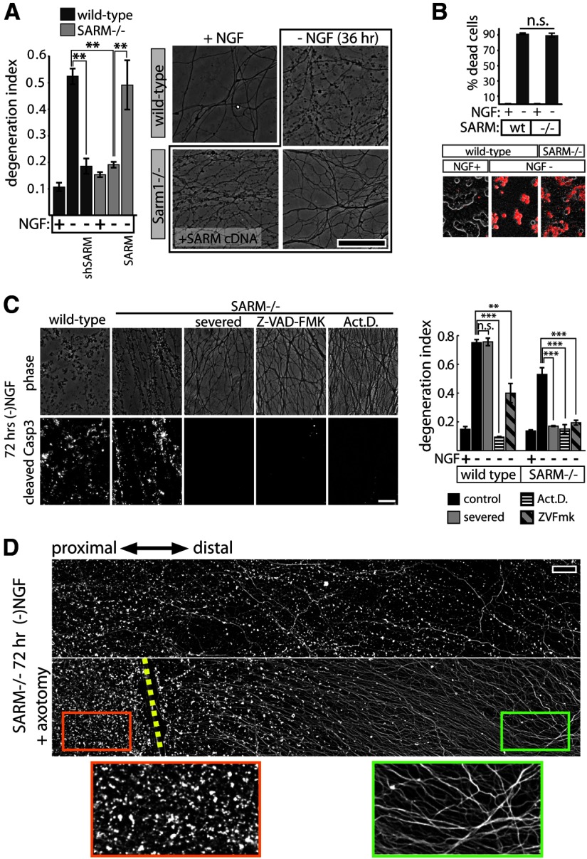
Figure 2.
SARM plays a role in axon degeneration elicited by trophic factor withdrawal. A, NGF deprivation elicits axon degeneration in wild-type neurons that is suppressed by SARM knockdown (shSARM). SARM−/− neurons show suppressed axon degeneration that is restored by expression of SARM. Representative phase-contrast images are shown at the right. B, SARM ablation does not affect apoptosis of DRG neurons measured after 24 h of NGF deprivation using ethidium homodimer (EH) exclusion assay. Top, Quantification of EH-positive cells shows no difference in cell death between genotypes. Bottom, Representative images of EH positivity (red stain). C, Following 72 h trophic factor withdrawal, SARM−/− axons exhibit fragmentation and cleaved caspase-3 (Casp3) immunoreactivity comparable to wild-type axons. Axon degeneration and Casp3 are blocked by severing the axons, treating with caspase inhibitor Z-VAD-FMK (10 μg/ml), or the transcriptional inhibitor actinomycin D (Act.D.; 1 μg/ml). Axon degeneration quantification is shown in the bar graph (right). D, Fluorescence (α-tubulin stain) montages showing axotomy-induced axon protection in SARM−/− DRGs deprived of NGF for 72 h. Cut site is indicated by yellow dashed line. **p < 0.01; ***p < 0.001; error bars = SEM, scale bars, 50 μm.

