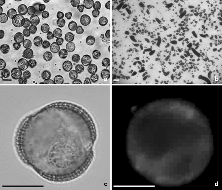
Fig. 1.
Microspore suspension of B. napus. Isolated microspore culture after 1 day at 32 ºC (a). Note that the average size of the majority of microspores is 20 μm in diameter. Microspore-derived embryos (black structures) of highly embryogenic ‘DH 4079’ line after 4 weeks of culture in NLN-13 medium on Petri dish (b). Uni-nucleate microspore after 1 day of heat shock treatment (c). Microspore after 1 day of heat shock treatment with incorporated fluorescent dye DPH. Blue fluorescence of DPH in UV light (d). Scale bar 10 μm (a), 1 mm (b), and 20 μm (c, d). Light microscope (a–c). Fluorescence microscope (d)

