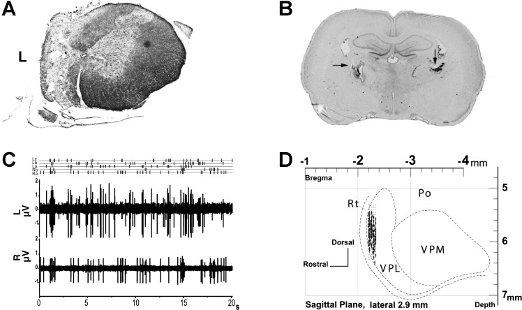
Fig. 1.
A: example of thoracic spinal cord hemisection (4 wk) at T10 (Nissl and myelin stain). B: bilateral electrolytic lesions in nucleus ventroposterior lateralis (VPL) recording sites (arrows). Intact preparation coronal section is shown. C: raw data in dual-channel recording (intact preparation). Three spikes sorted in multiunit recording from left VPL (La, Lb, and Lc) and 2 units sorted in recording from right VPL (Ra and Rb). D: VPL recording sites in sagittal plane for intact preparations. L, left; R, right; VPM, ventroposterior medial nucleus; Po, posterior nucleus; Rt, reticular nucleus.

