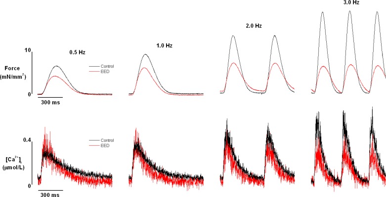
Fig. 2.
Raw recordings of force (top panels) and corresponding intracellular Ca2+ transients (bottom panels) of trabeculae before and after endocardial endothelium denudation (EED) with 0.1% Triton X-100. Trabeculae were superfused with Krebs-Henseleit solution and stimulated at the frequencies shown. Experimental conditions: external [Ca2+] = 0.5 mmol/l, temperature = 22°C.

