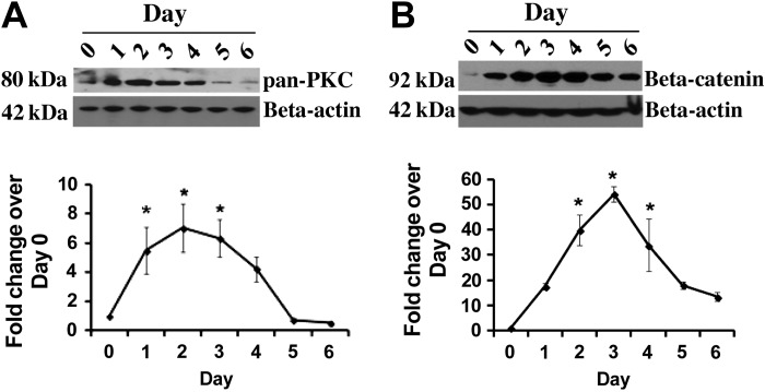
Fig. 4.
Activation of protein kinase C (PKC) and β-catenin during transdifferentiation. Representative immunoblots of pan-PKC and β-catenin from lysate from ATII cells cultured from 0 to 6 days are shown (A and B). β-Actin was immunoblotted as a loading control. Bottom: densitometry for pan-PKC (n = 4) and β-catenin (n = 3). *Significant difference from day 0 (P < 0.05).

