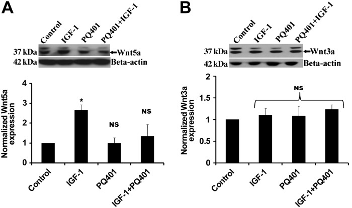
Fig. 5.
Wnt5a activation was stimulated by IGF-I. ATII cells (day 2) were treated with IGF-I (50 ng/ml), PQ401 (500 ng/ml), or the combination for 24 h, and lysates were collected. Representative immunoblots of Wnt5a and Wnt3a are shown (A and B) along with densitometry for Wnt5a (n = 4) and Wnt3a (n = 4). *Significant difference from untreated controls (P < 0.05). NS, values were not significant compared with control.

