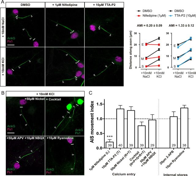
Figure 2.
Only activation of L-type calcium channels is essential for AIS relocation. A, Effects of L- and T-type channel block. Left, Sample images of DGCs treated with DMSO, 1 μm nifedipine, or 10 μm TTA-P2 in NaCl or KCl treatment groups. Right, Mean ± SEM of AIS start (S), maximum (M), and end (E) position for each treatment and subsequent calculation of AMI. B, Example images of DGCs treated with KCl in the presence of 50 μm nickel, cocktail of VGCC inhibitors, 50 μm APV + 10 μm NBQX, or 10 μm ryanodine. C, AMI mean ± SEM for each drug experiment. Brackets denote VGCC subtype blocked in each experiment. ***p < 0.0001, single-sample t test of AMI vs 1. Numbers within bars show the number of cells for each experiment. Scale bars, 10 μm. Thick white line denotes axon start, and white triangles illustrate AIS location. AnkG, Ankyrin-G; Px1, prox1.

