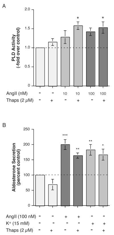
Figure 9.
Thapsigargin Enhanced the Effect of AngII on PLD Activation Cells but Had No Effect on AngII- (or Elevated [K+]e-) Stimulated Aldosterone Secretion in H295R. (A) H295R cells were pre-labeled for 20-24 hours with 2.5-5 μCi/mL [3H]oleate in serum-free medium. Cells were then stimulated with or without 10 or 100 nM AngII in the presence or absence of 2 μM thapsigargin (Thaps) or vehicle (0.2% DMSO) in the presence of 0.5% ethanol. Lipids were extracted and separated by thin-layer chromatography. Values represent the radioactivity in phosphatidylethanol relative to the control and are expressed as the means ± S.E.M. of 3 separate experiments; *p<0.05 versus the control value. (B) H295R cells were incubated for 5 hours with medium containing no additions (Con), 15 mM KCl (K+) or 100 nM AngII in the presence or absence of 2 μM thapsigargin (Thaps) or vehicle (0.1-0.2% DMSO) as indicated. Supernatants were collected and assayed for aldosterone secretion by radioimmunoassay. Values represent the means ± S.E.M. of 4-5 separate experiments; *p<0.05, **p<0.01, ***p<0.001 versus the control value.

