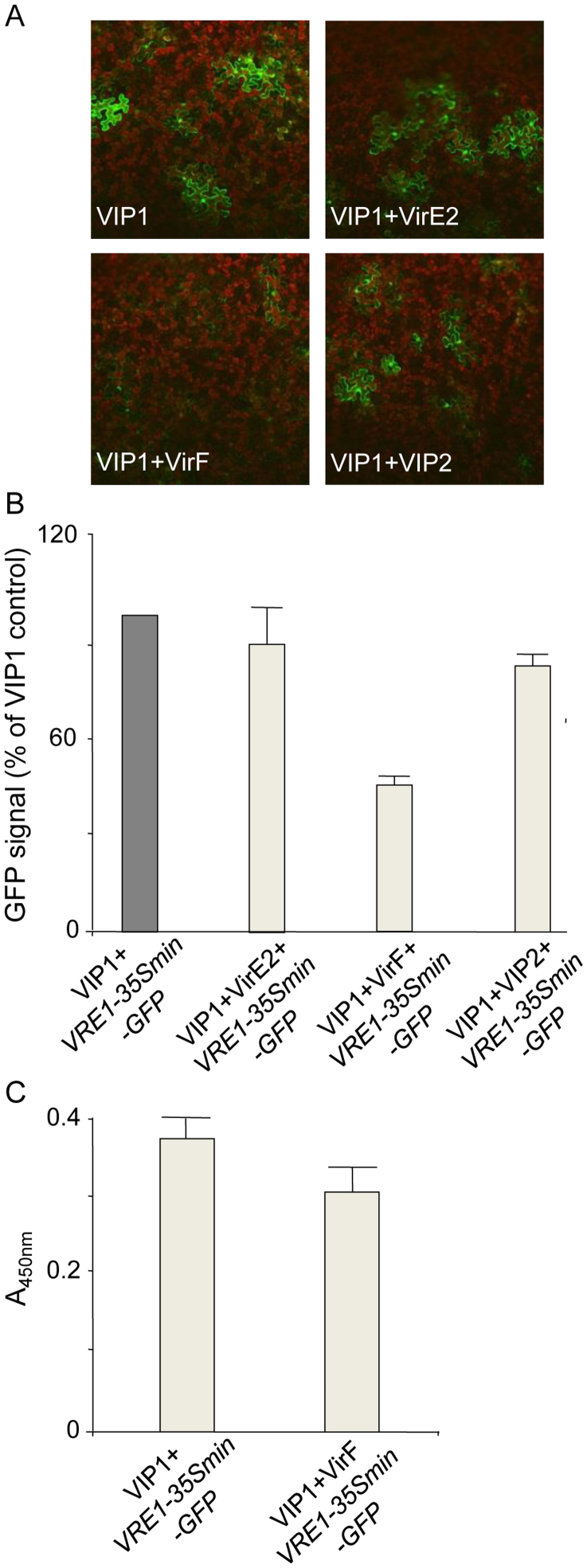
Figure 4. Effect of VIP1 interactors on promoter activation and VRE1 binding by VIP1.
(A) VIP1 activation of the transiently expressed VRE1-35Smin-GFP reporter in the presence of coexpressed VirE2, VirF, and VIP2. GFP is in green, plastid autofluorescence is in red. All images are single confocal sections. (B) quantification of VIP1-induced expression of VRE-35Smin-GFP in the presence of coexpressed VirE2, VirF, and VIP2. GFP signal was calculated as percent of the signal measured with the VRE1-35Smin-GFP reporter coexpressed with VIP1 without interactors, which was defined as 100% signal. All quantified data are shown as mean of three experiments with indicated standard deviations; standard deviation for measurements of the VRE1-35Smin-GFP reporter itself was 11.0%. (C) VIP1 binding to VRE1-35Smin-GFP is not affected by the presence of VirF.

