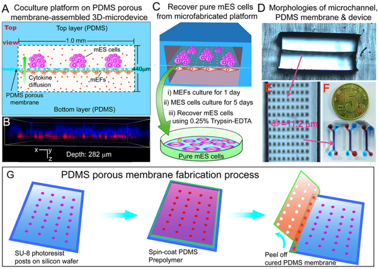
Figure 1. Schematic of PDMS porous membrane-assembled microfluidic co-culture platform.
(A) Top view illustration of the device dimensions and mES cells/mEFs co-culture on microchannels. (B) Confocal morphology showing the mES cells/mEFs coculture (depth 282 μm); stem cells layer strained with Hoechst 33342) (blue fluorescence) and feeder layer with RFP-mEFs. (C) Recover pure mES cells using 0.25% Tryspin-EDTA after 5 days culture. (D, E and F) Morphologies and structure of assembled microchannels, PDMS porous membrane (thickness: 10 μm; pore diameter: 11.2 μm) and fabricated microdevice. (G) Fabrication of PDMS porous membrane using standard soft lithography and replica molding techniques.

